Back to procedures
Procedure

Charcot Foot & Diabetic Foot Reconstruction in Hyderabad

Expert Charcot foot and diabetic foot reconstruction in Hyderabad. Explore treatment options, deformity correction, ulcer care and recovery at StepSure Clinic.

Charcot Foot & Diabetic Foot Reconstruction in Hyderabad

A foot wound that does not heal, a change in the shape of your foot, or sudden swelling in a person with diabetes should never be ignored. These problems can sometimes indicate Charcot foot or a serious diabetic foot complication that needs prompt medical attention.

At StepSure Foot & Ankle Clinic, Hyderabad, Dr. Uma Sai Manukonda, Consultant Foot & Ankle Orthopaedic Surgeon, evaluates foot and ankle deformities, Charcot neuroarthropathy, diabetic foot ulcers associated with structural problems, and conditions that may require reconstructive surgery.

Charcot foot develops when nerve damage reduces the ability to feel injuries and pressure. The bones and joints can become damaged, causing the arch to collapse or the foot to change shape. These changes may create areas of excessive pressure, leading to ulcers, infection and difficulty walking.

Not every patient with Charcot foot needs surgery. Early treatment often involves immobilisation, pressure relief and carefully selected protective footwear. When a severe deformity, instability, recurrent ulcer or bone prominence cannot be managed adequately with non-surgical treatment, reconstructive surgery may be considered.

The goal of Charcot and diabetic foot reconstruction is to create a more stable, functional and pressure-tolerant foot, help wounds heal where possible, and reduce the risk of further complications.

If you are looking for Charcot foot treatment or diabetic foot reconstruction in Hyderabad, an individual assessment can help identify the most appropriate treatment for your condition.

When Should You Seek Urgent Medical Care?

  • A newly hot, red or swollen foot: If you have diabetes or reduced foot sensation, seek urgent medical assessment, even if there is little or no pain. Suspected active Charcot foot requires prompt protection from further loading and specialist evaluation.
  • An open wound, pus, spreading redness or blackened skin: Seek urgent medical care. Fever, rapidly spreading infection or feeling severely unwell may indicate a limb-threatening or life-threatening infection.
1. What Is Charcot Foot and Diabetic Foot Reconstruction?

What Is Charcot Foot?

Charcot foot, also called Charcot neuroarthropathy, is a condition in which bones, joints and supporting tissues of the foot become damaged in the presence of nerve dysfunction. Diabetes is a common cause of the nerve damage associated with this condition.

When sensation is reduced, a person may continue walking on an injured foot without realising the extent of the damage. Repeated loading can lead to fractures, joint displacement and progressive changes in foot shape.

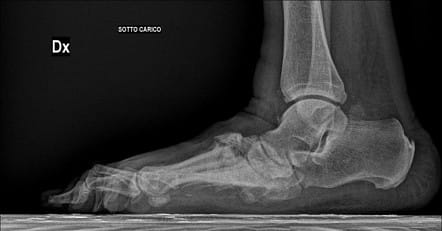
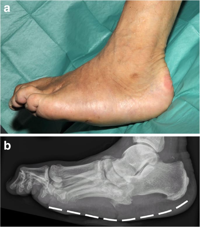
In advanced cases, the arch may collapse, producing a rounded or rocker-bottom shape. The abnormal shape can place excessive pressure on the skin beneath the foot.

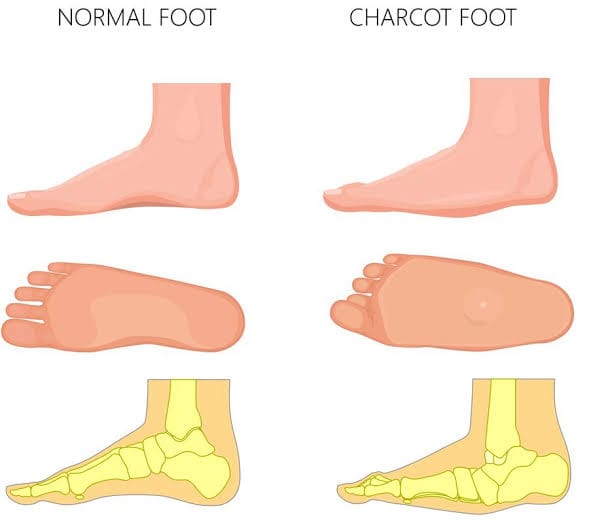
Comparison of a normal foot arch with arch collapse in Charcot foot.
Normal foot versus Charcot deformity

What Is Diabetic Foot Reconstruction?

Diabetic foot reconstruction is a group of surgical procedures used to correct selected foot deformities, improve stability, remove pressure-producing bone prominences, and address structural problems associated with ulcers.

Depending on the condition, reconstruction may involve:

  • Realigning bones and joints.
  • Fusing damaged or unstable joints.
  • Removing a bony prominence that repeatedly causes an ulcer.
  • Correcting tendon imbalance or associated deformity.
  • Treating infected or dead bone when indicated.
  • Reconstructing the foot after selected fractures or joint collapse.

The exact treatment depends on the stage of Charcot disease, skin condition, blood supply, presence or absence of infection, bone quality and overall medical fitness.

2. Who May Need Charcot or Diabetic Foot Reconstruction?

A consultation with a foot and ankle orthopaedic surgeon may be appropriate if you have:

  • A collapsed arch or major change in foot shape associated with diabetes.
  • Repeated ulcers over a bony prominence.
  • A deformity that makes it difficult to wear protective footwear.
  • An unstable foot or ankle that interferes with standing and walking.
  • A chronic ulcer associated with abnormal pressure or underlying bone deformity.
  • Bone infection requiring surgical treatment as part of a coordinated care plan.
  • A foot deformity that remains difficult to protect despite casting, bracing or custom footwear.
  • A previous Charcot reconstruction that has developed recurrent deformity or hardware-related problems.

A person with an early, active Charcot foot may need urgent immobilisation rather than immediate reconstructive surgery. Surgery is generally considered after weighing the expected benefits against the risks.

3. Common Symptoms of Charcot and Diabetic Foot Problems

Warmth, Redness and Swelling

One foot may be noticeably warmer or more swollen than the other. These symptoms can occur before obvious bone changes appear on X-rays.

Change in Foot Shape

The arch may flatten, the foot may widen, or the ankle may become unstable. Pain can be surprisingly mild when sensation is reduced.

A Wound That Does Not Heal

Pressure beneath a deformed foot may cause a persistent ulcer. An ulcer can become infected and, in some cases, lead to infection of the underlying bone.

Other symptoms include numbness, callus formation, difficulty fitting into shoes, repeated skin breakdown, and difficulty walking.

Important: Not every hot, swollen foot is Charcot foot. Infection, a fracture, gout and other conditions can produce similar symptoms. A proper examination is essential.

4. Causes and Conditions Treated

Diabetes-related peripheral neuropathy is a major risk factor for Charcot foot. Other nerve disorders can also cause the condition.

StepSure's Charcot and diabetic foot reconstruction service is intended for assessment and treatment planning for conditions such as:

  • Active and chronic Charcot neuroarthropathy.
  • Midfoot collapse and rocker-bottom deformity.
  • Hindfoot and ankle Charcot deformity.
  • Diabetic foot ulcers associated with abnormal pressure.
  • Prominent bones causing recurrent ulcers.
  • Selected cases of diabetic foot osteomyelitis, or infection of bone.
  • Deformity following fractures and joint dislocations.
  • Tendon imbalance contributing to abnormal loading.
  • Recurrent wounds associated with structural deformity.

The treatment plan may involve an orthopaedic surgeon, diabetes physician, vascular specialist, wound-care team, infectious disease specialist and orthotist, depending on the patient's needs.

5. When Is Surgery Recommended?

Surgery is not the first treatment for every Charcot foot. Many patients can be managed with appropriate immobilisation, offloading and long-term protective footwear.

Reconstructive surgery may be considered when:

  • A severe deformity cannot be accommodated safely in a brace or shoe.
  • A bony prominence causes recurrent ulceration.
  • The foot or ankle remains unstable.
  • There is a high risk of skin breakdown because of abnormal pressure.
  • A deformity prevents safe weight-bearing despite appropriate non-surgical care.
  • Infection or damaged bone requires surgical intervention.
  • Previous treatment has not achieved a foot that can be protected reliably.

The timing of surgery matters. In active Charcot disease, the initial priority is usually to protect the foot and control inflammation. Selected unstable or otherwise complicated cases may require earlier surgery. In chronic disease, reconstruction is often planned after careful assessment of the bone, skin and soft tissues.

The decision also considers circulation, infection, blood glucose control, nutritional status, kidney function, ability to follow postoperative restrictions and the patient's rehabilitation support.

6. Non-Surgical Treatment Options

Non-surgical care is essential both before and after reconstruction. It may also be the definitive treatment for many patients.

Immobilisation and Offloading

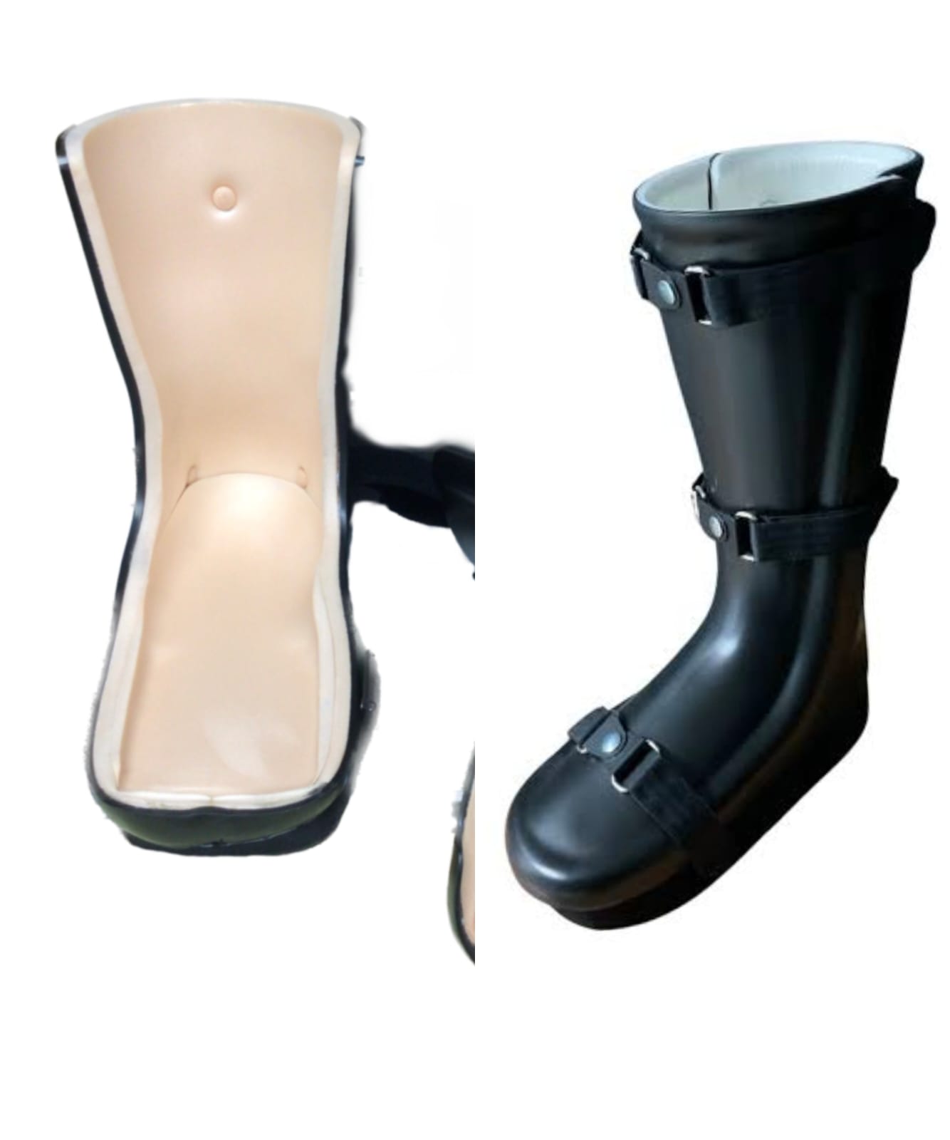
A carefully monitored cast, removable walker or other protective device reduces stress on the affected foot. A non-removable knee-high device may be appropriate for selected patients with active Charcot disease or certain ulcers, provided it is safe and suitable for their circumstances. The device and weight-bearing instructions must be individualised.

Rest and Activity Modification

Avoid unnecessary walking on a suspected active Charcot foot. Use crutches, a walker or a wheelchair if advised. Do not continue normal activity simply because the foot is not painful.

Custom Orthotics and Footwear

Once the foot is sufficiently stable, specially designed insoles, accommodative footwear or a custom brace may help distribute pressure and reduce ulcer risk.

Custom-moulded diabetic foot orthotic designed to reduce pressure on vulnerable areas.
Protective footwear and custom orthotics

Physiotherapy

Rehabilitation can help maintain strength, balance and mobility when appropriate. Weight-bearing exercises must not begin until the treating team confirms that they are safe.

Medications

Pain relief and treatment of associated medical conditions may be needed. Antibiotics are used when a bacterial infection is suspected or confirmed; they do not treat Charcot bone and joint damage itself.

Wound Care and Metabolic Control

Dressings, removal of unhealthy tissue when indicated, pressure relief, diabetes management, nutrition and circulation assessment all contribute to treatment.

Injections

Injections are not a standard treatment for active Charcot foot. They are considered only when there is a separate, clearly diagnosed indication and the clinician considers them appropriate.

Treatment recommendations follow the principles described in the International Working Group on the Diabetic Foot (IWGDF) guidance.

7. How Is Charcot Foot Reconstruction Performed?

There is no single operation suitable for every patient. The procedure is selected according to the location of the deformity, the condition of the skin, bone quality, stability and the presence or absence of infection.

Step 1: Clinical Assessment and Investigations

The surgeon examines the foot, skin, ulcers, sensation, circulation and alignment. Weight-bearing X-rays may be useful when safe and appropriate. CT scans can provide additional detail about bone structure, while MRI may help assess early bone changes or suspected infection.

Blood tests, vascular investigations and other medical assessments may be required.

Weight-bearing foot X-ray showing midfoot collapse associated with Charcot neuroarthropathy.
Diagnostic X-ray

Step 2: Prepare the Foot for Treatment

Before elective reconstruction, the team aims to control active infection, protect the skin, improve diabetes management and assess blood supply. Some patients need wound treatment, vascular intervention or staged surgery before definitive reconstruction.

Step 3: Correct the Deformity

Depending on the problem, the surgeon may perform one or more procedures:

  • Osteotomy: Cutting and repositioning a bone to correct alignment.
  • Arthrodesis: Fusing one or more joints to create a stable foot.
  • Exostectomy: Removing a prominent area of bone that causes pressure.
  • Tendon balancing: Adjusting selected tendons when muscle imbalance contributes to deformity.
  • Debridement: Removing infected or unhealthy tissue when required.
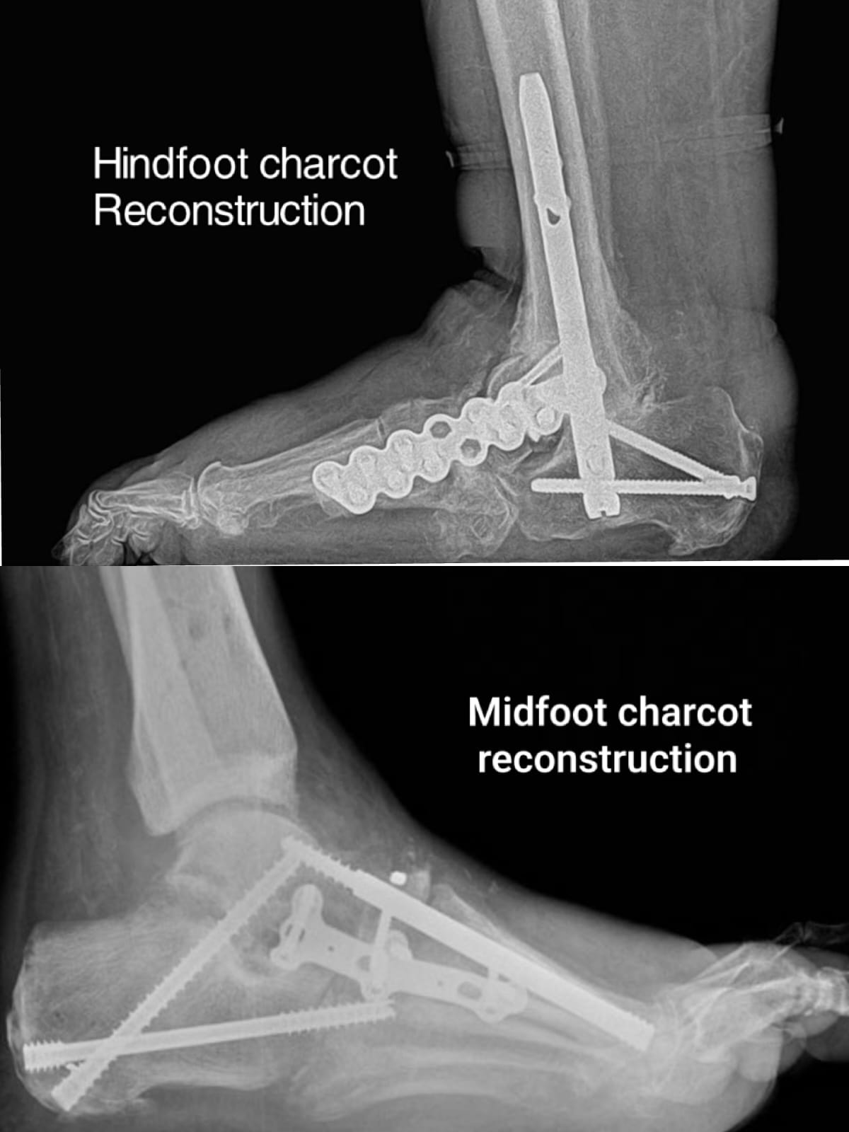
Illustration of bone realignment and internal fixation during Charcot foot reconstruction.
Reconstruction illustration

Step 4: Stabilise the Foot

Plates, screws, rods or other fixation devices may be used to hold the bones in the planned position. The fixation method depends on the bone involved, the extent of deformity and the quality of the bone.

Step 5: Protect the Reconstruction

After surgery, the foot is protected in a splint, cast or brace. The team monitors the incision, swelling, circulation, infection risk and bone healing.

Some patients require staged procedures rather than a single operation. The specific surgical plan and risks should be discussed before treatment.

8. Procedures That May Be Combined With Reconstruction

Depending on the patient's condition, the following procedures may be performed alone or together:

Midfoot Fusion
Stabilises damaged joints and corrects collapse of the arch.
Hindfoot or Ankle Fusion
May be required for severe hindfoot or ankle instability and deformity.
Corrective Osteotomy
Repositions bone to improve alignment and pressure distribution.
Exostectomy
Removes a bony prominence responsible for repeated skin pressure or ulceration in selected patients.
Achilles Tendon Lengthening or Tendon Balancing
May help reduce abnormal forefoot pressure in carefully selected cases.
Ulcer Debridement
Removes unhealthy tissue around a wound when indicated.
Bone Debridement and Infection Surgery
Removes infected or dead bone when required, with appropriate microbiological testing and antimicrobial treatment.
Soft-Tissue Coverage
Selected complex wounds may require specialist wound closure or reconstructive soft-tissue procedures.

Not every patient needs multiple procedures. Combining operations is considered only when the expected benefit justifies the additional surgical and healing risks.

9. Potential Benefits of Reconstruction

When appropriately selected, Charcot and diabetic foot reconstruction may help:

  • Improve foot and ankle alignment.
  • Increase stability for standing and walking.
  • Reduce pressure over vulnerable skin areas.
  • Help prevent recurrent ulcers caused by structural deformity.
  • Improve the ability to accommodate protective footwear.
  • Address selected cases of instability or infection-related bone damage.
  • Support limb preservation when a stable, protectable foot can be achieved.

Outcomes vary. Reconstruction cannot reverse nerve damage caused by diabetes, eliminate every risk of ulceration or guarantee that amputation will never be needed.

10. Recovery Timeline After Charcot Foot Reconstruction

Recovery is often longer than after routine foot surgery because diabetes, neuropathy, bone damage and poor circulation can delay healing. The following is a general guide, not a fixed schedule.

Weeks 1–2: Initial Healing and Protection

The foot is protected in a splint or cast. Wound checks, swelling control, blood glucose management and prevention of complications are priorities. Weight-bearing is restricted as instructed.

Weeks 2–6: Wound Monitoring and Follow-Up

The incision and skin are assessed, and X-rays may be obtained. Cast or brace adjustments may be needed. Many patients still require strict protection and limited or no weight-bearing.

Weeks 6–12: Assessment of Bone Healing

Imaging and clinical findings guide the next stage. Some patients can begin gradual, protected weight-bearing, while others need continued immobilisation.

Months 3–6: Progressive Rehabilitation

Patients who demonstrate adequate healing may gradually increase walking, transition to a suitable brace or custom footwear, and work on mobility and strength.

Months 6–12: Long-Term Recovery and Protection

Further improvement in walking function may occur. Some patients need longer to achieve bone fusion or wound healing and may require long-term bracing and surveillance.

These timeframes are approximate. Complex reconstructions may require several months of protected weight-bearing, and complete recovery can take a year or longer. Do not increase walking based on the calendar alone.

11. Physiotherapy and Rehabilitation

Rehabilitation is tailored to the stage of healing and the type of surgery.

Initially, the focus is on safe transfers, maintaining mobility in unaffected joints, preventing falls and learning how to use walking aids. Once the surgeon confirms that the reconstruction is sufficiently stable, a physiotherapist may introduce appropriate range-of-motion exercises, strengthening and gait training.

Patients also need guidance on safe footwear, daily skin inspection and long-term pressure management.

Because neuropathy may reduce pain as a warning signal, a painless foot is not necessarily a healed foot. New swelling, redness, blisters or skin breakdown should be assessed promptly.

12. Risks and Complications

Charcot and diabetic foot reconstruction can be complex. Potential complications include:

  • Wound-healing problems or wound breakdown.
  • Surgical-site infection or deep bone infection.
  • Delayed bone healing or failure of bones to fuse.
  • Fixation failure, loosening or breakage.
  • Recurrent deformity or continued pressure over the foot.
  • New ulcers caused by altered pressure distribution.
  • Blood clots, anaesthetic complications or other general surgical risks.
  • Persistent swelling, stiffness or difficulty walking.
  • Further surgery, including revision reconstruction or, in severe cases, amputation.

Risk is influenced by factors such as circulation, infection, blood glucose control, kidney disease, nutritional status, smoking, bone quality and the extent of reconstruction.

A careful preoperative assessment and long-term follow-up help identify and manage these risks. They cannot eliminate them entirely.

13. Expected Outcomes

The aim of reconstruction is to achieve a foot that is stable, appropriately aligned and easier to protect with suitable footwear or a brace.

Some patients regain useful walking ability and experience fewer pressure-related wounds. Others may continue to need walking aids, customised footwear or additional treatment.

The outcome depends on the initial deformity, circulation, infection status, bone quality, healing response and adherence to postoperative restrictions.

Even after successful reconstruction, diabetes-related neuropathy remains. Daily foot checks, appropriate footwear, diabetes management and regular follow-up are important for protecting the foot in the long term.

14. Why Choose StepSure Foot & Ankle Clinic?

StepSure Foot & Ankle Clinic focuses on the assessment and treatment of foot and ankle conditions, including complex deformities and selected diabetic foot problems.

Dedicated Foot & Ankle Practice

Foot and ankle conditions often require detailed assessment of alignment, joint stability, pressure distribution and footwear. A focused evaluation helps identify the problems that need treatment.

Personalised Treatment Planning

Treatment is based on the patient's symptoms, examination, imaging, circulation, skin condition and medical history. Surgery is considered when appropriate, rather than assumed to be necessary for every patient.

Modern Surgical Techniques

Where clinically appropriate, treatment may involve reconstructive fusion, corrective osteotomy, tendon procedures or minimally invasive techniques. The choice depends on the condition; minimally invasive surgery is not suitable for every complex deformity.

Imaging and Deformity Assessment

Weight-bearing X-rays, when safe and appropriate, can help assess foot alignment under load. Additional imaging may be needed for complex bone deformity or suspected infection.

Rehabilitation and Long-Term Protection

The treatment plan considers postoperative rehabilitation, protective footwear and long-term follow-up so that the reconstructed foot remains well protected over time.

Patients from Nizampet, Kukatpally, KPHB, Miyapur, Bachupally, Pragathi Nagar, Chandanagar, Hafeezpet, Madinaguda, Ameenpur, Suraram, Jeedimetla, Moosapet, Kondapur, Gachibowli, Hitech City, Jubilee Hills, Banjara Hills, Lingampally, Beeramguda, Kompally, Suchitra, Quthbullapur and Medchal can enquire about consultations and treatment planning.

If you are searching online for Charcot foot treatment in Hyderabad, diabetic foot reconstruction in Hyderabad or a foot and ankle orthopaedic surgeon in Hyderabad, seek an assessment that considers both the bone and joint deformity and the health of the skin and blood supply.

For a suspected active Charcot foot, a new ulcer or signs of infection, seek urgent medical evaluation rather than waiting for a routine appointment.

FAQs

Common patient questions

No. Many patients are managed with immobilisation, offloading and protective footwear. Surgery is generally considered when a severe deformity, instability, recurrent ulceration or bone prominence cannot be managed adequately with non-surgical treatment.
A newly hot, red or swollen foot, an open wound, pus, spreading redness or blackened skin, fever, or feeling severely unwell should be assessed urgently, even if there is little or no pain.
Depending on the condition, reconstruction may involve realigning bones (osteotomy), fusing damaged or unstable joints (arthrodesis), removing a prominent area of bone (exostectomy), tendon balancing, and removing infected or unhealthy tissue when required.
Recovery is often longer than after routine foot surgery. Early weeks focus on wound healing and protection, with gradual protected weight-bearing over the following months. Complex reconstructions can take a year or longer, and timelines vary between patients.
No. Reconstruction cannot reverse diabetic neuropathy. Daily foot checks, appropriate footwear, diabetes management and regular follow-up remain important for protecting the foot in the long term.
StepSure Foot & Ankle Clinic™Foot & Ankle Clinic

StepSure Foot & Ankle Clinic — Primary evening clinic in Nizampet, Hyderabad, with morning consultations at Jubilee Hills by appointment.

© 2026 Step Sure Clinic. All rights reserved.